Cyclin L1 (Isoform 1)抗体
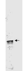
WB analysis of mouse brain tissue lysate using GTX48663 Cyclin L1 (Isoform 1) antibody.
Loading : 35 μg
Dilution : 1:3500

Western blot using GeneTex's Protein A Purified anti-Cyclin L1a antibody shows detection of a band ~59 kDa corresponding to a Cyclin L1a (arrowhead) present in mouse brain whole cell lysate. Approximately 35μg of lysate was separated by 4-20% SDS-PAGE followed by transfer to nitrocellulose. After blocking the membrane was probed with the primary antibody diluted to 1:3,500 for 2h at room temperature.
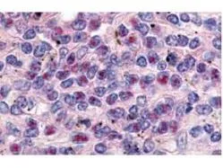
GeneTex's anti-Cyclin L1a antibody was used at a 10 μg/ml to detect Cyclin L1a in a variety of tissues including breast (collagen), heart, kidney (distal tubules), liver, skeletal muscle, ovary μgranulosa and oocyte), pancreas (islet), placenta (trophoblast), prostate (epithelium), skin, spleen (endothelium), stomach (chief), testes (seminiferous epithelium and leydig), thymus (Has-sal's corpuscle and lymphocytes) and uterus μglandular epithelium and stroma). Low to moderate background staining was noted. This image shows Cyclin L1a staining of human spleen. Tissue was formalin-fixed and paraffin embedded.
-
宿主Rabbit
-
克隆Polyclonal
-
同种型IgG
-
实验应用WB IHC-P IP ELISA
-
种属反应Human, Mouse, Drosophila