SCN1B抗体
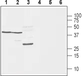
WB analysis of rat brain (lanes 1 and 4), mouse brain (lanes 2 and 5) and rat skeletal muscle (lanes 3 and 6) tissue lysates using GTX16973 SCN1B antibody preincubated with or without immunogen peptide.
Dilution : 1:400
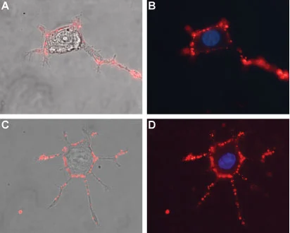
Live cell imaging analysis of live and intact differentiated PC12 cells using GTX16973 SCN1B antibody. PC12 differentiation was induced by Native mouse NGF 2.5S protein (>95%).
Red : Primary antibody
Blue : DAPI for nuclear staining
Dilution : 1:50
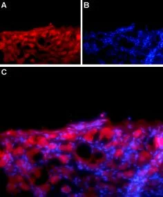
IHC-Fr analysis of rat DRG tissue using GTX16973 SCN1B antibody.
Panel A : NaVβ1 (red) appears in the cell bodies of the DRG neurons.
Panel B : Nuclear staining using DAPI (blue).
Panel C : Merged image of A and B.
-
宿主Rabbit
-
克隆Polyclonal
-
同种型IgG
-
实验应用WB ICC/IF IHC-Fr LCI
-
种属反应Mouse, Rat